Back to Course
Electrocardiology
0% Complete
0/0 Steps
-
Basics of ECG Interpretation11 Topics
-
Normal ECG Parameters
-
ECG Interpretation of Chamber Enlargement4 Topics
-
Dysrhythmias
-
Bradycardia
-
Heart Block3 Topics
-
Sick Sinus Syndrome
-
Tachycardia8 Topics
-
Hyperkalemia
-
Myocardial Hypoxia/Ischemia
-
Low Amplitude QRS Complex
-
Wide QRS Complex
-
Bundle Branch Block
-
Differentials for ECG Abnormalities
Lesson 3,
Topic 4
In Progress
Right Ventricular Enlargement
Lesson Progress
0% Complete
Dog:
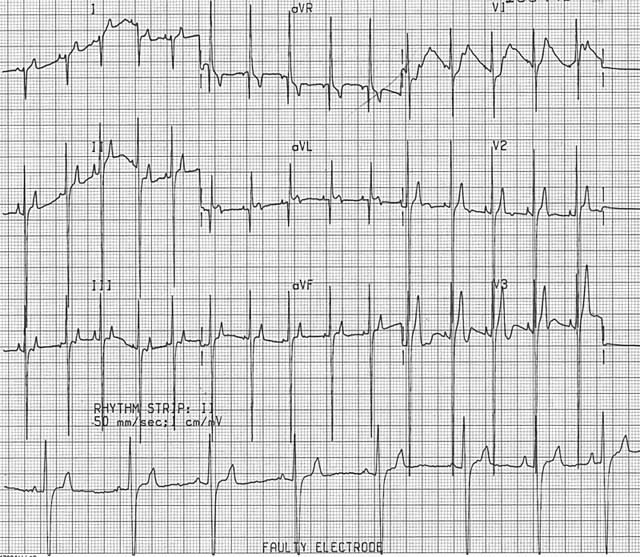
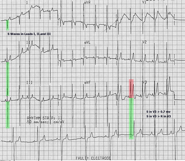
- Presence of an S wave in leads I, II, and III.
- MEA in the frontal plane shifted to the right (pointing to the right ventricle): 100o to – 75o
- Deep S wave in lead V3; S >= 0.7 mV
- S>R in lead V3
If all 4 criteria are present, there is strong evidence of right ventricular enlargement. The presence of 1 or 2 criteria is still strongly suggestive of right ventricular enlargement
Cat: MEA in the frontal plane shifted to the right (points to the right ventricle): 160° to -75°


