Back to Course
Electrocardiology
0% Complete
0/0 Steps
-
Basics of ECG Interpretation11 Topics
-
Normal ECG Parameters
-
ECG Interpretation of Chamber Enlargement4 Topics
-
Dysrhythmias
-
Bradycardia
-
Heart Block3 Topics
-
Sick Sinus Syndrome
-
Tachycardia8 Topics
-
Hyperkalemia
-
Myocardial Hypoxia/Ischemia
-
Low Amplitude QRS Complex
-
Wide QRS Complex
-
Bundle Branch Block
-
Differentials for ECG Abnormalities
Lesson Progress
0% Complete
ECG Findings:
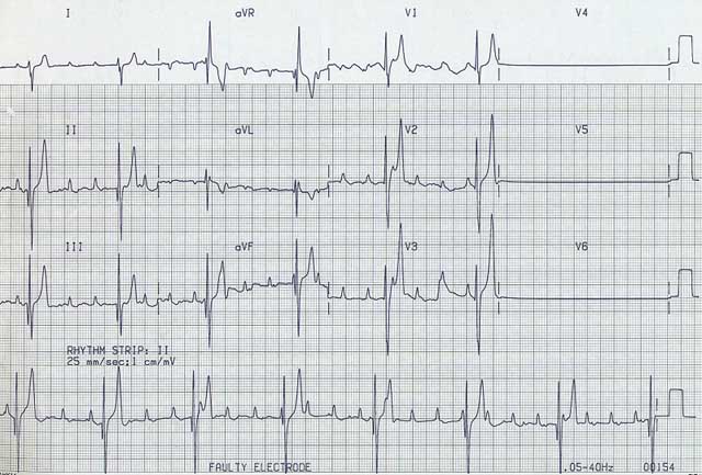
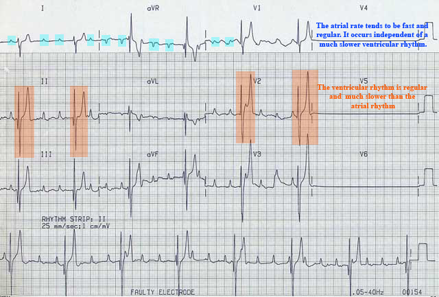
- The atrial rate and rhythm is independent of a much slower ventricular rhythm.
- The atrial rate tends to be fast and regular.
- The ventricular rhythm is regular and much slower than the atrial rhythm.
- Dog – ventricular rate approx. 40 – 60 bpm
- Cat – ventricular rate approx. 90 – 120 bpm
- The resultant ventricular activation is initiated from latent, subsidiary automatic pacemaker foci. The resultant rhythm is referred to as an escape rhythm. If the escape rhythm originates from the AV nodal area, the QRS morphology may look relatively normal. Usually, however, the escape rhythm originates from lower down and the QRS morphology tends to be abnormal (wide and bizarre).


Etiology:
- Due to the same etiologies as for 2nd degree Heart Block Mobitz Type 2
Consequences:
- Can cause all the signs of reduced cardiac output
Treatment:
- Atropine or glycopyrrolate are usually not effective
- IV isoproterenol may increase the ventricular escape rate
- Pacemaker therapy is usually the only effective therapy
