Cardiovascular Physiology and Pathophysiology
-
Physiology
Structure and Function4 Topics -
Lymphatics and Edema Formation
-
The Microcirculation
-
Vascular Control3 Topics
-
The Cardiac Cycle
-
Determinants of Myocardial Performance7 Topics
-
Neuro-Control of Heart and Vasculature4 Topics
-
Electro-Mechanical Association4 Topics
-
Electrical Side of the Heart4 Topics
-
PathophysiologyDefining Heart Failure
-
Causes of Heart Failure
-
MVO2 and Heart Failure
-
Cardiac Output and Heart Failure7 Topics
-
Compensation for Circulatory Failure
-
Vascular Tone in Heart Failure
Comparative Anatomy
Species differences in conduction system anatomy
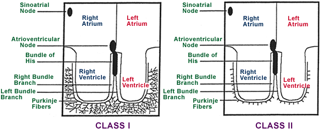
Based on the distribution of the Purkinje fibers, animals can be divided into two groups:
Class I: Hoofed mammals (horses, cows, sheep, pigs) and dolphins have a Purkinje fiber distribution that penetrates extensively from the endocardium to epicardium and also contains extensive cross fiber anastomotic bridges. The result is that the majority of the ventricular mass is activated simultaneously. The resultant QRS complex on the ECG represents primarily the activation of the base of the ventricular muscle. In general one cannot make a statement about ventricular mass based on the QRS morphology in these animals.
Class II: Dogs, cats, rats and people have a purkinje fiber distribution that penetrates minimally from the endocardium into the myocardium. The purkinje fibers do not penetrate more than 1/3rd of the distance from the endocardium to the epicardium. The result is that a wave of depolarization washes through the ventricular mass from the endocardium to the epicardium. The resultant QRS complex on ECG has a morphology that is determined by the mass of the ventricles in these animals. Hence, we can make a statement about ventricular enlargement based on the ECG.

Refer to electrocardiographic evaluation for more information on this topic.
